Catherine Sheehan, age 94, is admitted to the medical-surgical unit with a new diagnosis of heart failure and acute kidney injury. Laboratory results show an elevated B-type natriuretic peptide (BNP) level (above 20,000 pg/mL); blood urea nitrogen (BUN), creatinine, and all other levels are normal. On hospital day 3, her BUN, creatinine, and BNP levels decrease.
History and assessment hints
When Vanessa, the med-surg nurse, conducts her initial assessment at 7:30 PM, she finds Mrs. Sheehan awake, alert, and oriented. Her respiratory rate (RR) is regular at 18 breaths/minute; lung sounds are diminished in the lower fields. A spot-check peripheral oxygen saturation (SpO2) reading is 97% on 4 L oxygen/minute by nasal cannula.
When Vanessa checks on Mrs. Sheehan again at 10:30 PM, she finds her respirations labored and her RR increased to 24. Nonetheless, the patient says, “I’m breathing just fine.” Her SpO2 is 85% and her systolic pressure has climbed to 150 mm Hg.
When Vanessa notifies the physician of these findings, he orders supplemental oxygen titrated to an SpO2 of 92%, a STAT chest X-ray, 40 mg I.V. furosemide, bronchodilator protocol, and a ventilation/perfusion (V/Q) lung scan (which turns out to be negative for pulmonary embolism).
To achieve an SpO2 of 92%, Vanessa must place Mrs. Sheehan on a nonrebreather mask. Vanessa checks every 15 to 20 minutes for changes in her condition. At midnight, she finds the patient asleep, with an RR of 20 and SpO2 of 93%.
On the scene
At 1:00 AM, Vanessa notes Mrs. Sheehan’s breathing is shallow and rapid (28), her breath sounds are diminished throughout, and her SpO2 is 95%. Also, she is disoriented to place and time. Concerned that Mrs. Sheehan’s respiratory pattern has changed again despite her normal SpO2, Vanessa calls the physician and obtains an arterial blood gas (ABG) sample, as ordered. ABG results are pH 7.27, PaCO2 75 mmHg, PaO2 251 mmHg, and HCO3– 33 mEq/L.
Clearly, Mrs. Sheehan has adequate oxygenation, but her elevated PaCO2 signifies inadequate ventilation, indicating she’s in acute respiratory failure (ARF). She is transferred to the intensive care unit (ICU).
Outcome
In the ICU, Mrs. Sheehan receives diuretic therapy, dobutamine to improve cardiac contractility, and continuous bilevel positive airway pressure (BiPAP) for 24 hours. Then she’s weaned to oxygen therapy via nasal cannula and transferred back to the med-surg unit. Two days later, she’s discharged to a nursing home.
Education and follow-up
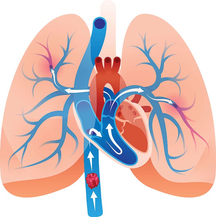
In ARF, gas exchange deteriorates. Factors that increase ARF risk include chronic obstructive pulmonary disease and pulmonary edema. ARF can be hypoxemic or hypercapneic. Mrs. Sheehan initially had hypoxemic failure from heart failure, then later developed acute hypercapneic ARF from a ventilation problem involving CO2 retention.
BiPAP, the preferred noninvasive ventilatory support method for ARF patients, has two pressure settings—inspiratory positive airway pressure (IPAP) and expiratory positive airway pressure (EPAP). IPAP assists gas exchange, aiding ventilation. EPAP prevents alveolar collapse and increases oxygen driving pressure to support oxygenation. BiPAP promotes gas exchange and allows respiratory recovery by decreasing breathing workload in patients with hypercapneic RF.
As Mrs. Sheehan’s experience shows, you can’t rely solely on SpO2 when evaluating a patient’s breathing adequacy. Instead, obtain a full set of vital signs and check for slight changes in the respiratory rate, pattern, and depth by assessing respirations for a full minute. Remember—O2 saturation reflects oxygenation status, not ventilatory status.
References
Boldrini R, Fasano L, Nava S. Noninvasive mechanical ventilation. Curr Opin Crit Care. 2012;18(1):48-53.
Johnson CL, Anderson MA, Hill PD. Comparison of pulse oximetry measures in a healthy population. Medsurg Nurs. 2012;21(2):70-6.
Kraynek B, Best J. What are the clinical indications for noninvasive positive pressure ventilation? Hospitalist. December 1, 2011. www.the-hospitalist.org/article/what-are-the-clinical-indications-for-noninvasive-positive-pressure-ventilation/.
Lewis SL, Dirksen SR, Heitkemper MM, Bucher L. Medical-Surgical Nursing: Assessment and Management of Clinical Problems. 9th ed. St. Louis: Mosby; 2013.
Sinuff T. Noninvasive positive pressure ventilation for acute respiratory failure: What role is there for patients declining intubation or choosing palliation? Prog Palliat Care. 2011;19(5), 223-9.
Soo Hoo GW. Noninvasive ventilation. http://emedicine.medscape.com/article/304235-overview
The authors work at Trinity Mother Frances Hospitals and Clinics in Tyler, Texas. Janette Gasaway is a staff nurse in the emergency department. When this article was written, Julie Miller was the staff development educator in critical care.