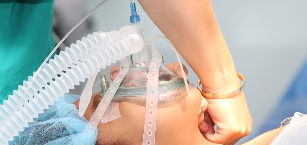
It’s your worst nightmare: At 8 A.M., you walk into the room of Laura M, a 35-year-old patient who had surgery yesterday, for your morning assessment. She’s unresponsive. The night nurse told you in report that she’d given Laura morphine I.V. every 2 hours for postoperative pain; since then, she said, Laura had been sleeping quietly, waking only during hourly rounds to ask for more pain medication. When the patient-care technician last checked vital signs at 6 A.M., Laura’s respiratory rate was 16 breaths/minute and oxygen saturation (Sao2) 98% on room air.
Faced with an unresponsive patient, you decide to act on the standing order for naloxone. You call out for someone to get the drug as you assess Laura’s pulse and respirations. She still has a pulse but her respiratory rate is just six breaths/minute. Your colleague begins ventilations with a bag-valve mask as you slowly administer naloxone until Laura begins to wake up. When her respiratory rate starts to improve, you heave a huge sigh of relief while your charge nurse arranges for her transfer to the intensive care unit (ICU). Crisis averted.
At the end of your shift, you check on Laura in the ICU. Her nurse tells you Laura has been complaining of poorly controlled pain throughout the day, but the physician is reluctant to order more opioids because of her near-miss this morning.
This scenario strikes fear in the hearts of bedside nurses. Opioid-induced respiratory depression (OIRD) is a life-threatening complication of opioid analgesia. Even the most opioid-tolerant patient isn’t immune. Respiratory arrest is a contributing factor in about one-third of the 750,000 cardiopulmonary arrests that occur annually in U.S. hospitals, and about half of these patients had received an opioid. Mortality for in-hospital cardiopulmonary arrest may be as high as 80%, with the worst outcomes occurring on medical-surgical units. Between 2006 and 2009, postoperative respiratory failure was the third most common safety incident in American hospitals, affecting an estimated 600,000 patients yearly at a cost of $1.5 billion.
Professional associations and regulatory agencies have called for action repeatedly to prevent OIRD in the hospital setting. In 2012, The Joint Commission (TJC) issued an alert urging hospitals to take action to increase opioid safety and reduce opioid-related sentinel events. The Anesthesia Patient Safety Foundation (APSF) has called for OIRD to be designated a “never” event. In 2011, the American Society for Pain Management Nursing (ASPMN) issued evidence-based guidelines to help nurses monitor for OIRD in acute-care settings.
Nurses bear the brunt of responsibility for monitoring hospital patients for OIRD, yet we have little control over the medication regimens they’re receiving. This is where capnography can play a lifesaving role. This noninvasive technique detects early signs of OIRD by measuring exhaled carbon dioxide (CO2). Early detection promotes timely rescue, particularly on med-surg units where nurse-to-patient ratios are higher and critical events are less likely to be witnessed. Numerous studies show capnography detects signs of respiratory depression earlier and more effectively than visual respiratory assessments or pulse oximetry. This crucial tool can help you keep patients safe.
Understanding OIRD
Defined as a decrease in baseline ventilatory function after opioid administration, OIRD manifests as a respiratory rate between eight and 10 breaths/minute, oxygen saturation as measured by pulse oximetry (Spo2) below 90%, and end-tidal carbon dioxide (ETco2) above 50 mm Hg. Severe respiratory depression occurs when the respiratory rate falls below eight breaths/minute with Spo2 below 85% for at least 6 minutes.
But these thresholds aren’t exact, and OIRD has occurred even when vital signs are outside these parameters. So always treat the patient, not the numbers. Keep in mind that sedation, a common opioid side effect, precedes respiratory depression. Be sure to assess regularly for sedation (along with respirations and pain) and document sedation using a validated scale, such as the Pasero Opioid-Induced Sedation Scale.
When OIRD is detected early, nonpharmacologic interventions may be effective in preventing the condition from worsening. Upright patient positioning, talking to or shaking the patient gently, and encouraging breathing may be enough to arouse the patient and stimulate breathing.
When OIRD isn’t detected early, naloxone must be given to reverse opioid respiratory depressant effects. Yet when administered improperly, naloxone reverses opioid analgesic effects, leading to severe pain and distress for your patient. Naloxone acts quickly but doesn’t last long, so patients may need multiple doses, depending on duration of the opioids they received. About 0.2% to 0.7% of patients receiving postoperative opioids require naloxone rescue. In the United States, this equates to about 20,000 patients every year.
Although naloxone was once thought to cause only minor side effects (such as nausea and vomiting from increased pain), we now know it’s far from benign. It may lead pulmonary edema, arrhythmias, hypertension, and cardiac arrest. (See Giving naloxone to adults with OIRD.)
Who’s at risk for OIRD
All patients are susceptible to OIRD—whether opioid-naïve or tolerant, whether they’re receiving an oral-only opioid regimen or an I.V. infusion, whether the nurse administers the opioid or the patient uses a patient-controlled analgesia (PCA) device. But certain factors can raise the risk. ASPMN has identified the following high-risk groups: patients with renal dysfunction, chronic obstructive pulmonary disease, heart failure, obstructive sleep apnea (OSA), or obesity.
In OSA, the upper airway becomes occluded during sleep. The condition increases OIRD risk because opioids may relax pharyngeal tone and worsen airway occlusion. OSA prevalence may be as high as 5%, although only about 15% of sufferers have a confirmed diagnosis. The high rate of undiagnosed OSA means clinicians may not recognize a patient’s risk. A body mass index above 30 and a neck circumference of 17.5″ or more indirectly indicate OSA and are considered OIRD risk factors. Never ignore loud snoring—an ominous sign of airway obstruction.
Certain iatrogenic factors also increase risk. According to TJC, 11% of opioid-related sentinel events from 2004 to 2011 stemmed from excessive dosing, drug interactions, and adverse effects. Patients receiving both benzodiazapines and opioids or continuous opioid infusions and those within the first 24 hours after surgery involving general anesthesia are at greater risk for OIRD. What’s more, OIRD can occur not just with continuous opioid infusions (including continuous infusion mode on PCA) but also with intermittent parenteral opioid injections. One analysis of OIRD cases over a 1-year period found only 20% involved continuous opioid infusions; the rest were linked to intermittent I.V. bolus injections or oral opioids (or concurrent use of both oral and parenteral opioids).
This finding helps us understand where patients at high risk for OIRD tend to be located in hospital settings: Generally, continuous opioid drips are given on critical care units, where continuous monitoring is standard practice. In contrast, RN-administered intermittent parenteral opioids and oral opioids usually are given on med-surg units, where only intermittent bedside monitoring of respiratory status is the norm.
Multiple factors can influence whether a patient is more or less likely to experience OIRD. Patients who become opioid tolerant from long-term opioid therapy at home for chronic pain may require significantly higher opioid doses for acute pain in the hospital. (See How opioids ease pain and affect respiration.) Although chronic opioid use at consistent daily doses may provide some protection against OIRD, dose escalation can put the patient at risk. One study showed a higher OIRD incidence among opioid-tolerant patients than opioid-naïve patients. Also, use of illegal opioids, such as heroin, leads to opioid tolerance and creates added challenges for pain control and OIRD risk in hospitals. (See OIRD risk factors.)
Comparing capnography and pulse oximetry
Capnography and pulse oximetry measure two different physiologic processes—ventilation and oxygenation, respectively.
- Capnography measures ETco2, which reflects ventilation—an indicator of how well the patient is managing the mechanics of breathing.
- Pulse oximetry reflects oxygen saturation of the blood—how much oxygen is getting into the blood after passing through the lungs. A low Spo2 can alert you to a ventilation problem, but only because that problem has caused an oxygenation problem. In other words, low Spo2 is a relatively late OIRD indicator.
In monitoring patients for OIRD, pulse oximetry is no substitute for capnography—especially in patients receiving supplemental oxygen. Although the additional oxygen boosts Spo2, it does so artificially, potentially masking compromised ventilation. Relying on pulse oximetry can be misleading and even dangerous, giving a false sense of security that your patient is ventilating well. Studies have validated that capnography is more effective than pulse oximetry in detecting early signs of OIRD in patients receiving supplemental oxygen. (See Pulse oximetry vs. capnography in OIRD monitoring.)
Capnography in action
In capnography, a nasal cannula captures exhaled breath and measures ETco2. Most capnography devices provide both a waveform and a digital reading of ETco2, usually displayed in mm Hg. The target range is 35 to 45 mm Hg; higher levels indicate worsening ventilation. Capnography also measures apneic events (pauses in breathing lasting more than 10 seconds), as well as respiratory rate.
Placing the capnography cannula on the patient’s face is akin to initiating a nasal cannula for supplemental oxygen. Breath samples are obtained through both nostrils, and oxygen may be delivered simultaneously through small pinholes. Some types of tubing have extensions resembling a spoon in front of the mouth, which can be used to obtain readings if the patient breathes through the mouth instead of the nose (as often occurs in patients with sleep apnea).
But don’t rely solely on spot checks or short-term continuous ETCO2 readings. Also observe ETco2 trends over the course of your shift and the previous shift. Say, for instance, your patient’s ETco2 level has been hovering around 45 mm Hg for most of your shift. But if on the night shift he was maintaining 40 mm Hg and yesterday evening he was at 35 mm Hg, this shows an increasing trend that may signify a problem.
Many capnography devices can be integrated with PCA. With these systems, if the respiratory rate drops below the programmed parameter, the PCA shuts down and the patient can’t self-administer more opioid medication. This setup commonly is used with basal rates (continuous infusions) on PCA pumps, as continuous opioid infusion carries a greater risk. However, even incremental-only PCA regimens carry some risk, especially in the immediate postoperative period.
Typical capnography settings
Anesthesiologists have long relied on capnography in the operating room to monitor lung ventilation and help identify low cardiac output and pulmonary emboli. For many surgical procedures, capnography is mandatory for financial reimbursement. In postanesthesia care units, nurses use capnography to monitor patients during the immediate postoperative period when pain management can be challenging and OIRD risk is high. Nurses on critical care units use capnography to verify endotracheal and nasogastric tube placement, as well as for continuous monitoring of ventilation.
During procedural sedation, physicians, nurse anesthetists, and nurses rely on capnography to monitor ventilatory function. Physicians and nurse practitioners use capnography during cardiopulmonary resuscitation (CPR) to confirm endotracheal tube placement, assess CPR quality, and obtain early indication of return of spontaneous circulation.
Increasingly, nurses are using capnography on general med-surg units to monitor for OIRD, although this is still a novel approach in most hospitals. A national survey of nurses’ monitoring practices to prevent OIRD, published by ASPMN in 2013, revealed that pulse oximetry was widely used in the 99 facilities that responded. Only 2.2% used capnography for patients undergoing epidural therapy (but this rose to 6% for high-risk patients) and 1.5% used it for patients with PCA devices. Of the 23 responding facilities that used continuous capnography, 22 found it useful in detecting OIRD. No facilities routinely used capnography based on overall OIRD risk assessment, regardless of opioid administration method. Also, 75% of nurse respondents said they didn’t have continuous capnography devices available.
OIRD can arise quickly, and nurses may not always recognize it at an early stage. In critical care units, the higher level of general monitoring and lower nurse-to-patient ratios mean nurses are more likely to detect and recognize OIRD early. But postoperative patients commonly are transferred to general med-surg units, where nurses have more patients to care for and less time to spend with each patient. If OIRD occurs, they’re less likely to witness it immediately, so rapid response team activation may be delayed. Continuous monitoring and early detection with capnography makes this method a potentially valuable tool to help nurses ensure patient safety. However, most nurses are less familiar with capnography than pulse oximetry.
Who should be monitored with continuous capnography?
In 2011, APSF recommended continuous electronic monitoring for all postoperative patients receiving opioids. Also in 2011, the ASPMN Expert Consensus Panel published recommendations for nurses on monitoring for opioid-induced sedation and respiratory depression. These included individualizing the frequency, intensity, duration, and nature of monitoring (assessments of sedation levels and respiratory status, plus technology-supported monitoring) based on the type of opioid therapy, patient and iatrogenic risk factors, response to treatment, and facility policies. The panel stated that technology-supported monitoring (such as continuous pulse oximetry and capnography) can be effective for patients at high risk of OIRD based on thorough assessment of the risk factors described above.
So how do we implement these recommendations in day-to-day practice, especially on general med-surg units where seemingly every patient has some combination of known risk factors? Assessing individual risk for each patient receiving opioids is an unreliable, inconsistent way to predict OIRD. Yet electronic monitoring for every hospital patient receiving opioids isn’t feasible. What’s more, technology used for continuous capnography isn’t perfect; condensation in the tubing can cause false alarms, contributing to alarm fatigue for patients and nurses. As a result, nurses may lower preset thresholds or patients may refuse to wear the cannula. Until the technology is perfected, we must do the best we can to apply the monitoring to patients who need it most.
Capnography can be lifesaving for patients who clearly are at higher OIRD risk, such as:
- those who are within 24 hours postoperative, regardless of the amount of opioids they’re receiving and by what route
- those with confirmed or suspected OSA
- those on continuous opioid infusions
- opioid-naïve patients during their first 24 hours on opioids
- opioid-tolerant patients who’ve had a dose escalation in the previous 24 hours.
Also, patients receiving supplemental oxygen should be monitored with capnography whenever possible, because administered oxygen can mask hypoventilation.
For high-risk patients, continuous capnography monitoring is better than intermittent monitoring. Even if nurses conduct respiratory assessments every hour for a full 5 minutes, that leaves patients unmonitored 92% of the time. And studies show nurses and patient-care technicians don’t consistently conduct and document respiratory assessments. Although technological monitoring is no substitute for frequent face-to-face bedside assessment by a nurse, it can be a valuable supplement.
A shared responsibility
All hospital staff share the responsibility of keeping patients safe. OIRD is a problem of both overprescribing and undermonitoring. Nurses must advocate for sensible multimodal pain medications from the provider, whether that provider is a physician, physician’s assistant, or nurse practitioner.
Some prescribers may underdose opioids for fear of causing OIRD. But undertreated pain leads to poorer recovery, longer hospital stays, and low patient satisfaction—and jeopardizes the patient’s trust in the nurse. Availability of continuous capnography reassures clinicians that an extra layer of monitoring is in place to detect early signs of OIRD.
At the other end of the spectrum, prescribers may rely too heavily on high-dose opioid-only therapy and thus neglect to order nonopioids, adjuvant medications, or both. Range orders for opioids (for example, 2 to 4 mg morphine q 4 hours p.r.n. for moderate to severe pain) allow nurses to start low and go slow, which helps prevent OIRD. Capnography provides added security.
With proper education, patients and family members also can watch for signs and symptoms of respiratory depression, and many are savvy to capnography’s benefits. Public awareness and interest in capnography is growing. The “Promise to Amanda” website (www.promisetoamanda.org), set up by parents of a teenager who died of OIRD while in the hospital for a minor infection, is devoted to promoting capnography for all patients using PCA. The Physician-Patient Alliance for Health and Safety advocates spreading capnography use among physicians and the public.
Patient education
Another key component of OIRD prevention is patient education regarding the goals of pain management and the need to balance comfort with safety. The goal is to make patients comfortable enough to perform the functions necessary for recovery but not necessarily for completely eradicating pain, which might require high doses that could harm them. Patients benefit from information about expectations of pain control in the context of personal safety; this needn’t negatively impact patient satisfaction surveys. When patients believe hospital staff have done their best to address their pain even if they can’t eliminate it entirely, they report satisfaction with the care they received.
Revisiting the scenario
If Laura had been monitored with capnography, both you and she would have been alerted that her exhaled CO2 levels were climbing and her respiratory rate was slowing. The alarm might have triggered Laura to take deeper breaths, while alerting you to adjust her morphine dose. You would have requested orders for nonopioid pain medication and adjuvant medications for multimodal pain management. As a result, the prescriber wouldn’t have withheld Laura’s pain medications as a knee-jerk reaction, and Laura wouldn’t have suffered poor pain control all day. Her transfer to the ICU wouldn’t have been needed, and most likely she would have been discharged home within a day or two.
References
Bauman M, Cosgrove C. Understanding end-tidal CO2 monitoring. Am Nurse Today. 2012;7(11):12-7.
Dahan A, Aarts L, Smith TW. Incidence, reversal, and prevention of opioid-induced respiratory depression. Anesthesiology. 2010;112(1):226-38.
Jarzyna D, Jungquist CR, Pasero C, et al.; American Society for Pain Management Nursing. American Society for Pain Management Nursing guidelines on monitoring of opioid-induced sedation and respiratory depression. Pain Manag Nurs. 2011;12(3):118-45.
Jungquist CR, Karan S, Perlis ML. Risk factors for opioid-induced excessive respiratory depression. Pain Manag Nurs. 2011;12(3):180-7.
Kodali BS. Capnography outside the operating rooms. Anesthesiology. 2013;118(1):192-201.
Nisbet AT, Mooney-Cotter F. Comparison of selected sedation scales for reporting opioid-induced sedation assessment. Pain Manag Nurs. 2009;10(3):154-64.
Overdyk FJ, DeVita MA, Pasero C. Postoperative opioid-induced respiratory depression: current challenges and new developments in patient monitoring. Anesthes News. 2012;1-8.
Overdyk FJ, Guerra JJ. Improving outcomes in med-surg patients with opioid induced respiratory depression. Am Nurse Today. 2011;6(11):26-30.
Pasero C. The perianesthesia nurse’s role in the prevention of opioid-related sentinel events. J Perianesth Nurs. 2013;28(1):31-7.
Pasero C, McCaffery M. Pain Assessment and Pharmacologic Management. St. Louis, MO: Mosby; 2011.
The Joint Commission. Safe use of opioids in hospitals. Sentinel Event Alert. 2012;49;1-5.
Weinger MB, Lee LA. No patient shall be harmed by opioid-induced respiratory depression. Proceedings of “Essential Monitoring Strategies to Detect Clinically Significant Drug-Induced Respiratory Depression in the Postoperative Period” Conference. APSF Newsletter. 2011;26(2):21-40.
Whitaker DK. Time for capnography—everywhere. Anaesthesia. 2011;66(7):544-9.
Willens JS, Jungquist CR, Cohen A, Polomano R. ASPMN Survey – Nurses’ practice patterns related to monitoring and preventing respiratory depression. Pain Manag Nurs. 2013;14(1):60-5.
Heather Carlisle is a nurse practitioner at the University of Arizona Medical Center in Tucson and a clinical assistant professor in the College of Nursing at the University of Arizona. She is board certified in pain management by the American Society for Pain Management Nursing.


